
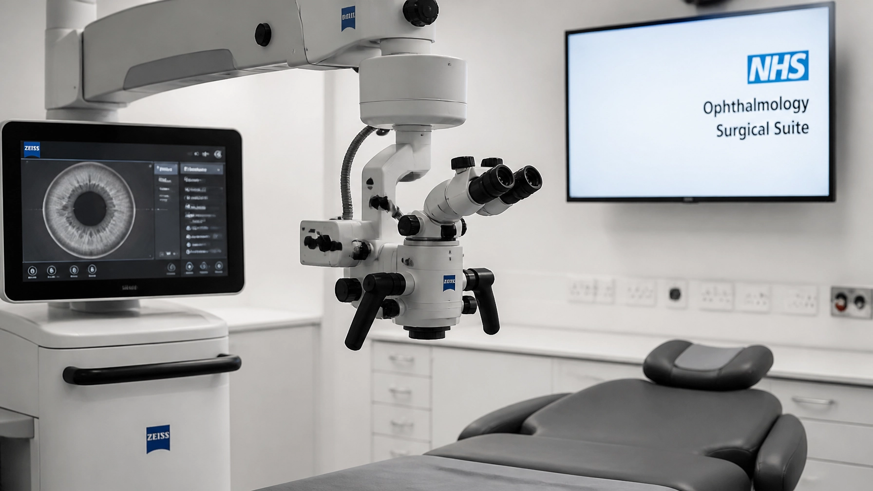

From Anatomy to Action: Why Foundational Science Drives Surgical Precision
Have you ever wondered why some surgeons move through a complex procedure with an almost telepathic ease? Why, in a crisis, their hands seem to know the path before their conscious mind has even finished processing the complication? It isn't just "talent." It isn't just "luck." It is the invisible architecture of foundational science.
In this instalment of our 'Surgery & Shift' series, inspired by the clinical precision of Miss Tina Parmar, we are stripping back the digital layers of modern ophthalmology to look at the raw anatomy beneath. In an era where we are surrounded by femtosecond lasers, robotic-assisted platforms, and AI-driven diagnostics, it is easy to fall into the trap of thinking the machine is the master.
It is not.
The machine is a tool. The anatomy is the map. If you don't know the map, the most expensive GPS in the world won't save you when the road gets rough. Let's have a look at why deep anatomical knowledge remains the ultimate secret to surgical success.
The Oculoplastic Blueprint: More Than Just Skin Deep
When we talk about oculoplastics, particularly in the context of Miss Tina Parmar’s expertise, we are discussing one of the most anatomically dense regions of the human body. The eyelid is not just a flap of skin. It is a sophisticated, multi-layered machine designed for one primary purpose: the protection of the ocular surface.

If you are a clinician looking to advance into surgical assisting or specialised co-management, you must understand the "Lamellar Concept." The division between the anterior lamella (skin and orbicularis muscle) and the posterior lamella (tarsal plate and conjunctiva) is the holy grail of eyelid reconstruction.
Why does this matter for precision? Because even a one-millimetre error in the positioning of the levator aponeurosis during a ptosis repair can be the difference between a perfect functional result and a lifelong corneal exposure issue. True surgical precision in oculoplastics isn't about how fast you can suture; it is about knowing exactly which anatomical plane you are in at every single second.
The key here is to recognise that anatomy is dynamic. It isn't a static drawing in a textbook. It is the way the septum interacts with the pre-aponeurotic fat pads. It is the tension of the medial and lateral canthal tendons. When you understand the "Why" behind the structure, the "How" of the surgery becomes second nature.
Refractive Precision as Structural Engineering
We often frame refractive surgery as a matter of physics and optics. We talk about dioptres, ablation depths, and wavefront patterns. But at its core, refractive surgery is structural engineering on a microscopic scale.
Whether it is a LASIK flap, a SMILE lenticule, or a refractive lens exchange, you are altering the biomechanical integrity of the eye. Foundational science tells us that the cornea is not a uniform piece of plastic. It is a living, breathing collagenous matrix with a highly specific arrangement of lamellae that provide its strength and transparency.
If you ignore the micro-anatomy: if you fail to account for the corneal nerves or the depth of the basement membrane: you aren't performing surgery; you are just operating a machine. Precision comes from knowing exactly how the eye will respond to that structural change. Will the epithelium thicken in response to the ablation? How will the biomechanical tension of the stroma shift?
Miss Tina Parmar’s focus on foundational science reminds us that refractive success is measured in years, not weeks. It is about the long-term stability of the eye's anatomy after we have finished our work.
The Fallacy of the Automated Eye
Let’s have a bit of tough love. There is a growing trend in the industry to rely on the "black box" of technology. We see candidates who can recite the technical specifications of a laser but cannot describe the layers of the tear film or the precise attachments of the extraocular muscles.
It’s easy to think, "The laser is accurate to the micron, so I don't need to worry."
Wrong.
The laser doesn't know if the patient has underlying thyroid eye disease that will affect their lid position post-surgery. The laser doesn't know if the patient's corneal endothelium is compromised. Foundational science is your safety net. It is what allows you to look at a "perfect" scan and say, "Something doesn't feel right here."
We hope for your success, but we also want you to be prepared for the worst-case scenario. When the power goes out, or the machine glitches, or the anatomy doesn't look like the textbook, your deep understanding of foundational science is all you have left.

Building Your Clinical Foundation: A Script for Success
If you are an optometrist or a pre-reg student looking to make a bigger impact in your role: perhaps even eyeing a move into a "mini-hospital" environment or a surgical centre: you need to change your approach. Stop reading the "User Manual" and start reading the "Anatomy Manual."
Here is a simple affirmation you can use during your next clinical session:
"I am not just measuring a surface; I am observing a biological system. My precision is rooted in my understanding of the structures I cannot see."
Let’s have a look at three actionable steps to boost your anatomical precision:
- Peer Review Every Case: Don't just tick the boxes. Join our Clinical Exchange forum and discuss the anatomical "Why" behind your complex cases.
- Visualise the Layers: When you look through the slit lamp, don't just see "the cornea." Visualise the epithelium, the stroma, and the endothelium. See the anatomical layers as distinct entities.
- Study the Surgical Path: Even if you aren't a surgeon, learn the surgical steps for the procedures you co-manage. Understanding where a surgeon makes an incision or places a suture will transform the way you handle post-operative care.
The Future of 'Surgery & Shift'
We are incredibly proud to highlight the work of pioneers like Miss Tina Parmar. Her commitment to foundational science is what sets a world-class surgeon apart from a technician. At NeedAnOptom Healthcare, we are not just filling positions; we are connecting clinical excellence with the platforms it deserves.

Whether you are a practice looking for surgical-grade talent or a candidate ready to take the next step in your career, we are here to guide you. Our mission is to ensure that the UK optical sector remains a world leader in clinical precision, supported by the best minds and the most robust foundational knowledge.
Finally, we must acknowledge that our digital home and the infrastructure that allows us to share these clinical insights are expertly managed by the team at ZeroPoint Creative. Their technical development ensures that our "Architecture of Care" is as stable and precise as the surgeries we describe.
Are you ready to move from anatomy to action? Search our latest clinical roles or sign up for job alerts today. Your next clinical shift is waiting.
NeedAnOptom Healthcare
Run by opticians, for opticians.
leave your comment